Posts

Showing posts from March, 2023

Instrumentation 6

Microscopy is the study of objects or samples that are too small to be seen by the naked eye. There are several types of microscopy, each with its own advantages and limitations. Here are the main types of microscopy: 1. Optical microscopy: This is the most common type of microscopy, which uses visible light to illuminate a sample. Optical microscopy can be further divided into several subtypes, such as brightfield, darkfield, phase contrast, fluorescence, and confocal microscopy. Optical microscopy is a technique that uses visible light to observe the sample under a microscope. It consists of several components, including an objective lens, an eyepiece lens, and a light source. The working of optical microscopy involves the following steps. The sample to be viewed is prepared by fixing it onto a glass slide and adding a stain or dye to enhance its contrast. The light source, located beneath the sample, emits light that is directed through the condenser lens to focus the light o

Branches of microbiology.

Image
Microbiology is a vast field that encompasses a variety of branches, each with its own specific focus and applications. Some of the major branches of microbiology are:   Bacteriology: This branch focuses on the study of bacteria, including their structure, function, and role in disease. Bacteriology is the branch of microbiology that focuses on the study of bacteria, which are single-celled microorganisms that can be found in a wide range of environments, including soil, water, and living organisms. Bacteriology involves the study of the morphology, physiology, biochemistry, genetics, and ecology of bacteria, as well as their interactions with other microorganisms and with their environment.   Bacteriologists study a wide range of topics related to bacteria, including their classification, taxonomy, and evolution, as well as their role in human health and disease. Bacteriology plays a critical role in the fields of medicine, public health, and agriculture, where it is used t

HOW/ WHERE/ WHY…. The SDS-PAGE Electrophoresis Explanation.

Image
INFORMATION: SDS-PAGE (Sodium Dodecyl Sulfate Polyacrylamide Gel Electrophoresis) is a widely used technique for separating and analyzing proteins. In this technique, proteins are first denatured and coated with anionic detergent SDS which gives all proteins a negative charge relative to their size. The negatively charged proteins are then separated by electrophoresis based on their size, as they move through a polyacrylamide gel matrix under an electric field. The polyacrylamide gel matrix has pores of different sizes, with smaller pores towards the bottom, allowing smaller proteins to move more quickly through the gel than larger ones. The separated proteins are then stained with a dye such as Coomassie Brilliant Blue or Silver Nitrate, which binds to the proteins, making them visible for analysis. The resulting pattern of separated proteins, known as a protein profile, can be used to identify and quantify individual proteins, as well as determine their molecular weight and relat

Citric Acid Cycle Explanation.

Image
The citric acid cycle , also known as the Krebs cycle or the tricarboxylic acid (TCA) cycle , is a series of biochemical reactions that occur in the mitochondria of eukaryotic cells and the cytosol of prokaryotic cells. The cycle plays a crucial role in the cellular respiration process, which generates energy in the form of ATP.   The cycle starts with the conversion of pyruvate, which is produced by glycolysis, into acetyl-CoA. This reaction is catalyzed by a complex of enzymes called the pyruvate dehydrogenase complex. During this reaction, one molecule of CO2 and two electrons are removed from pyruvate, which is then bound to Coenzyme A (CoA) to form acetyl-CoA.   Once acetyl-CoA enters the citric acid cycle, it combines with a four-carbon molecule called oxaloacetate to form a six-carbon molecule called citrate. This reaction is catalyzed by the enzyme citrate synthase. Citrate is then converted to isocitrate through a series of reactions catalyzed by aconitase and isocitr

The cell cycle

Image
The cell cycle is the process by which a single cell divides into two genetically identical daughter cells. It is a series of events that takes place in a cell leading to its division and duplication. The cell cycle is divided into two main stages: interphase and the mitotic phase.   Interphase: Interphase is the period between cell divisions. During this stage, the cell grows, replicates its DNA, and prepares for cell division. Interphase is divided into three phases:   G1 phase: During the G1 phase, the cell grows and synthesizes RNA and proteins needed for DNA replication.   S phase: During the S phase, DNA replication occurs, resulting in two identical copies of the genetic material.   G2 phase: During the G2 phase, the cell prepares for cell division, synthesizes microtubules, and checks the integrity of DNA. Mitotic Phase:   The mitotic phase is the period during which the cell divides into two daughter cells. The mitotic phase is divided into four stages

Recombinant DNA technology

Image
Recombinant DNA technology involves the manipulation of DNA molecules from different sources to create new DNA molecules. The most common vectors used in recombinant DNA technology are plasmids, bacteriophages, and artificial chromosomes. Plasmids are circular pieces of DNA found in bacteria and are commonly used as vectors in genetic engineering. They can be easily replicated and can carry foreign DNA into a host cell. Bacteriophages are viruses that infect bacteria, and they can also be used as vectors in genetic engineering. They can be modified to carry foreign DNA into a host cell, where it can be integrated into the host's genome. Artificial chromosomes are man-made DNA molecules that can carry large pieces of foreign DNA. They are designed to replicate and segregate like natural chromosomes and can be used to study the structure and function of complex genomes. Therefore, there are several vectors available for recombinant DNA technology, but plasmids, bacteriophages

DNA Sequencing Protocol

Image
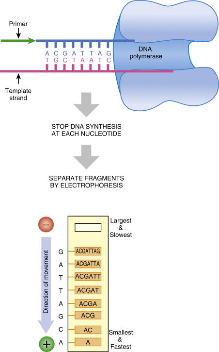
Step-by-step protocol for DNA sequencing:   DNA Extraction: The first step in DNA sequencing is to extract the DNA from the sample you wish to sequence. There are many DNA extraction protocols available depending on the sample type. Fragmentation: Next, the extracted DNA is broken down into smaller fragments using various techniques such as sonication or enzymatic digestion.   Library preparation:  These DNA fragments are then ligated to specialized adapters that allow them to bind to the sequencing platform. This process is called library preparation.   PCR amplification:  Once the DNA fragments have been ligated to the adapters, PCR (polymerase chain reaction) is used to amplify the number of fragments. This step is critical to obtain enough DNA for sequencing. Quality Control:   The quality of the DNA library is assessed using different methods such as gel electrophoresis or bioanalyzer.   Sequencing:  The sequencing platform then reads the DNA sequences. Diffe

DNA sequencing

Image
DNA sequencing is the process of determining the precise order of nucleotides (A, C, G, and T) in a DNA molecule. It is a fundamental technique in modern molecular biology and genetics that enables researchers to understand the genetic information that encodes an organism's traits, functions, and evolutionary history. The DNA sequencing process involves several steps, which are generally divided into three main stages: library preparation, sequencing, and data analysis. Here's an overview of each stage:   Library preparation: This stage involves extracting DNA from a biological sample (e.g., blood, tissue, or saliva), fragmenting it into smaller pieces, and attaching small DNA adapters to each fragment. The adapters serve as anchors for the sequencing machinery and allow for the DNA fragments to be amplified and sequenced. Once the DNA fragments have been prepared, they are loaded onto a sequencing instrument.   Sequencing: This stage involves using a sequencing ins

RNA Vs DNA

Image
  Note: Although RNA is usually single-stranded, it can sometimes form temporary double-stranded regions through base pairing within the molecule or with complementary RNA or DNA molecules.

Different types of DNA.

Image
DNA (Deoxyribonucleic acid) is a complex molecule that carries genetic information in living organisms. DNA is made up of building blocks called nucleotides, which are composed of a sugar molecule, a phosphate group, and a nitrogenous base. The four nitrogenous bases that makeup DNA are adenine (A), thymine (T), guanine (G), and cytosine (C) . The order of these bases, known as the DNA sequence, determines the genetic information encoded in DNA. There are different types of DNA, including: B-DNA:  B-DNA is the most common form of DNA, which is found in cells during normal physiological conditions. It is a right-handed double helix with ten base pairs per helical turn. The nitrogenous bases are stacked on top of each other, and the sugar-phosphate backbone twists around the axis of the helix. B-DNA is stable and is not easily disrupted by changes in temperature, pH, or salt concentration. A-DNA:  A-DNA is a right-handed double helix with 11 base pair

RNA Extraction

Image
RNA Extraction: RNA extraction is the process of isolating RNA from biological samples such as cells, tissues, or blood. RNA extraction is an important step in many molecular biology techniques such as gene expression analysis, RNA sequencing, and PCR. Materials needed for RNA extraction include: Biological sample (cells, tissues, blood, etc.) RNase-free tubes, pipettes, and tips RNase-free water or buffer RNA extraction kit or reagents Denaturants such as guanidine isothiocyanate or phenol Alcohol such as ethanol or isopropanol Buffers for lysis and washing Protease inhibitors to prevent protein degradation (optional) Equipment such as a centrifuge, vortex mixer, and thermocycler Gloves and lab coat to prevent RNase contamination Chemical preparation for RNA extraction depends on the specific RNA extraction method and kit used. Most RNA extraction kits provide pre-made buffers and reagents that are optimized for RNA isolation. However, for some methods, the following steps may be nece

Popular posts from this blog

what is Biostatistics.

Golden rice

STD 12th/Ch-Reproduction /Asexual reproduction & sexual reproduction